

中字视频见同名微博微信哟~~
翻译:雪球滚滚;校对:ivlvn;
词群:quizlet.com/_5nkskm
肺
主要功能:气体交换,吸入氧气和排出二氧化碳。
The main job of lungs is gas exchange, pulling oxygen into the body and getting rid of carbon dioxide.
正常情况下,吸气时,膈膜收缩向下移动,胸部肌肉收缩扩大胸腔 ==> 肺部压强变小,有助于吸入空气。呼气时,胸部肌肉放松,肺部恢复正常形状,排出空气。
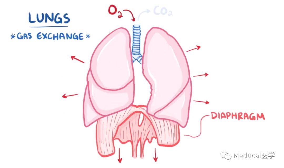
Normally, during an inhale: the diaphragm contracts to pull downward and chest muscles contract to pull open the chest, which helps suck in air like a vacuum, and then during an exhale: the muscles relax, allowing the lungs to spring back to their normal size pulling the air out.
鼻 腔
吸气时,气流通过鼻孔进入鼻腔,鼻腔中有大量分泌粘液的细胞。这种粘液又咸又稠,含有溶菌酶(可杀死细菌)。在鼻腔入口处的鼻毛表面覆盖着这种粘液,它们会吸附大的颗粒、花粉和细菌,形成鼻屎!
When you breathe in, air flows through the nostrils and enters the nasal cavity which is lined by cells that release mucus. That mucus is salty, sticky, and contains lysozymes, which are enzymes that help kill bacteria. Nose hairs at the entrance of the nasal cavity get coated with that mucus and are able to trap large particles of dust and pollen as well as bacteria, forming tiny dumps of boogers.
鼻旁窦
鼻腔和四个鼻窦(位于鼻腔周围含气的骨质腔)相连,分别是额窦、筛窦、蝶窦、上颌窦。 鼻旁窦协助调节吸入的空气,使它们温暖湿润。鼻旁窦还有共鸣作用,放大你的声音。这就是为什么当人感冒时,鼻子被粘液堵住,声音就会听起来异样。
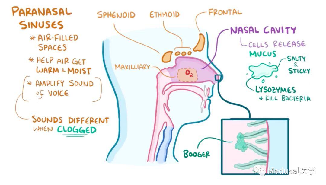
The nasal cavity is connected to four sinuses which are air-filled spaces inside the bones that surround the nose, there’s the frontal, ethmoid, sphenoid, and maxillary sinus. The paranasal sinuses help the inspired air to circulate for a bit so it has time to get warm and moist. The paranasal sinuses also act like tiny echo-chambers that help amplify the sound of your voice, which is you sound so different when they’re clogged with mucus during a cold!
咽 部
因此,从鼻腔进入咽部的空气是相对干净、温暖湿润的,连接这两者的区域叫做鼻咽部,连接咽部和口腔的部分叫做口咽部。软腭位于硬腭后方(用舌头能感受到)和像钟摆的悬雍垂悬挂在软腭的后方,它们一起形成一个挡板,挡住鼻咽,在人进食时防止食物掉入鼻咽部。 最后,这个部分是咽喉,咽部与喉咙相连的地方。
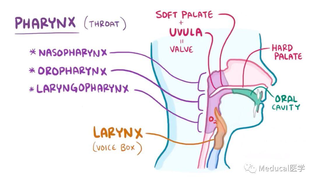
So the relatively clean, warm, and moist air goes from the nasal cavity into the pharynx or throat, the region connecting the two is called the nasopharynx, and the part connecting the pharynx to the oral cavity is called the oropharynx. The soft palate, the softer portion of the roof of your mouth behind the hard part that you can feel with your tongue, and the pendulum-like uvula hanging at its end move together to form a flap or valve that closes the nasopharynx off when you eat to prevent food from going up into the nasopharynx. Finally, there is the laryngopharynx, the part of the pharynx that’s continuous with the larynx or the voice box.
会 厌
空气和食物共享一条通路。但喉的顶部有一个匙状的软骨瓣,叫做会厌,它的作用就像一个盖子,当你吃东西的时候封住了气道,所以食物只能从食道进入胃。只要不是空气进入喉部,人就会将异物咳出体外。
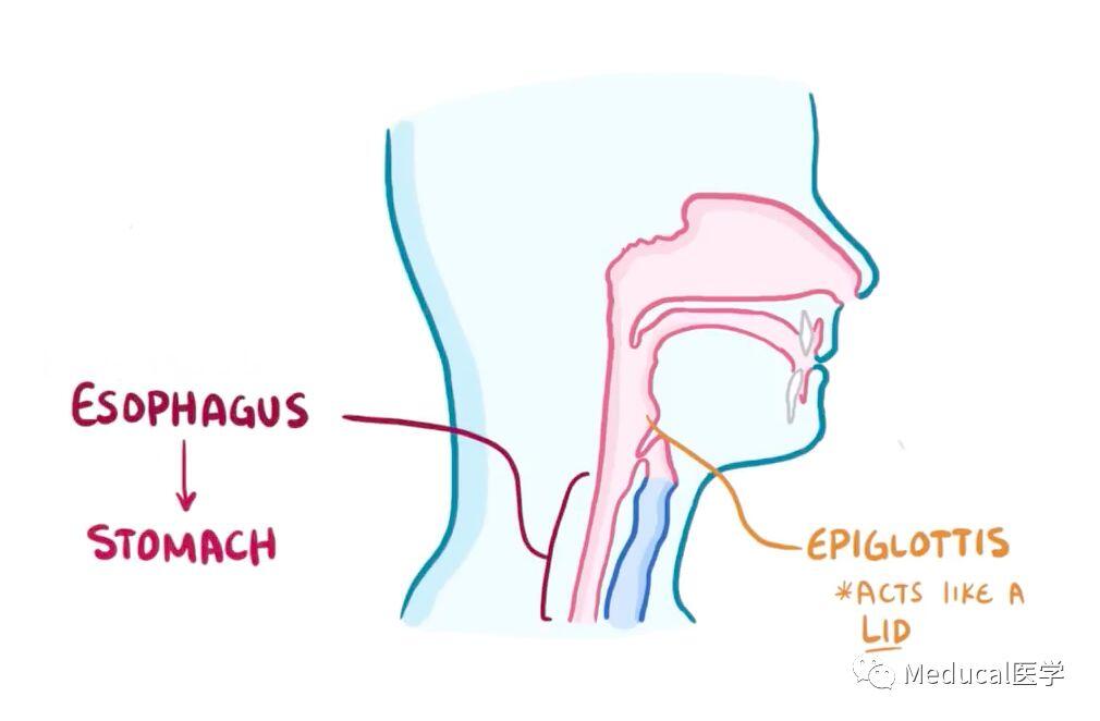
Up to this point, food and air share a common path. But the top of the larynx sits a spoon-shaped flap of cartilage called the epiglottis which acts like a lid that seals the airway off when you are eating, so that the food can only go one way – down the esophagus and towards the stomach. If anything other than air enters the larynx, then there’s a cough relax to kick it right out.
气 管
一旦空气进入咽喉,接下来就是气管,延伸为两侧的支气管。分叉处叫气管叉。然后接着看肺部,右肺有三叶:上叶、中叶和下叶。左肺只有两叶,即上叶和下叶。右侧支气管比左侧支气管较宽、更垂直,这就是为什么当人不小心吸入大颗粒物体无法咳出时,比如一颗花生,更容易掉入右肺而不是左肺的原因。支气管会反复分支成小支气管。气管和前三级支气管都非常宽大,有气管软骨作支撑。
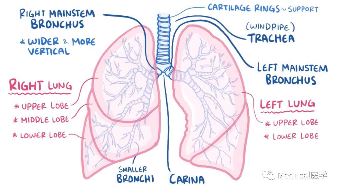
Now, once air makes it’s way into the larynx, it then continues down as the trachea or the windpipe, which splits into the two mainstem bronchi. The point at which they splits it called the carina.They then enters the lungs, and the right lung has three lobes-upper lobe, middle lobe, and lower lobe, and the left lung has just an upper lobe and lower lobe. The right mainstem bronchus is wider and more vertical than the left which is why if you accidently inhale something big that can’t get coughed out like a peanut, then it’s more likely to go into the right lung than the left. The mainstem bronchi then divided into smaller and smaller bronchi. The trachea and the first three generations of bronchi, are all pretty wide and use cartilage rings for support.
气管横断面
气管外围有一层平滑肌,里面含有神经,该神经属于自主神经系统。自主神经系统由两种基本神经模式组成:交感神经,即“战或逃”模式,就像远离一只火鸡;副交感神经,即“休息和消化”模式,就像在海滩上吃冰淇淋。
位于气管和初级支气管的分支的平滑肌上都有β2受体。再来看看火鸡这个例子,当人在奔跑时,交感神经系统激动β2受体,使气道扩张。但是这些气道也有M胆碱受体;副交感神经也可以激动它们,使气道收缩。
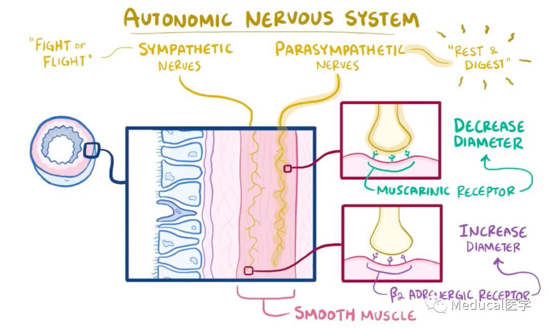
Taking a look at a cross section chunk, there’s also a layer of smooth muscle which has nerves of the autonomic nervous system within it. The autonomic nervous system is made up of two basic types of nerves: sympathetic nerves which are involved in “fight or flight” mode like running from a turkey and parasympathetic nerves which are involved in the “rest and digest” mode - like eating ice cream on the beach. Smooth muscle along the trachea and the first few branches of bronchi have beta 2 adrenergic receptors. Going back to that turkey, when you’re running, the sympathetic stimulate those beta 2 adrenergic receptors and increase the diameter of the airways. But those same airways also have muscarinic receptors which can get stimulated by parasympathetic nerves, causing a decrease in the diameter of airways.
大的气道内壁有大量纤毛柱状上皮细胞和少数分泌粘液的杯状细胞,这些细胞因看起来像玻璃杯而得名,其粘液吸附颗粒,然后纤毛柱状上皮细胞的节律摆动移动粘液和任何来自空气的颗粒进入喉部,可使颗粒被排出或被吞噬,这个机制叫粘液纤毛活动梯。
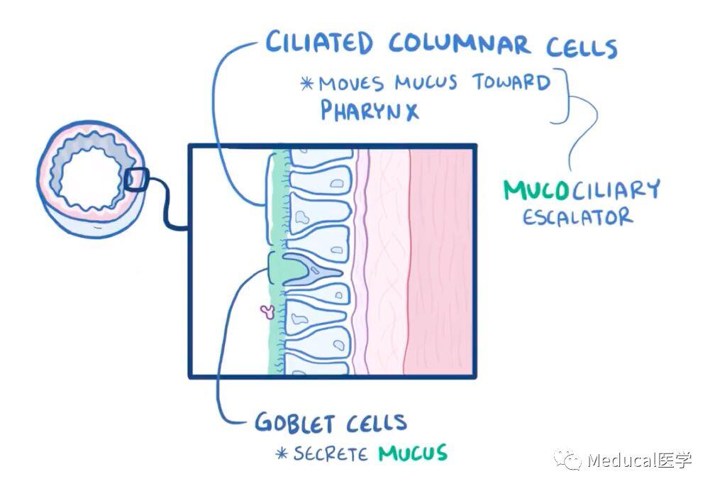
The large airways are lined mostly by ciliated columnar cells and a handful of goblet cells which get their name from looking like a wine goblet or glass, and secrete mucus. That mucus helps trap particles, and then the ciliated columnar cells beat rhythmically where they can either be spit out or swallowed this mechanism is known as the mucociliary escalator.
传导性细支气管
在经历三级支气管后,气道变得更狭窄,称为细支气管,这些气管可以保持开放而不需要软骨。空气经过越来越小的细支气管传导大约15-20代/级,它们统称为传导性细支气管,其管壁上也有纤毛柱状细胞和分泌粘液的杯状细胞,还有棒状细胞(可能分泌糖胺聚糖来保护细支气管内膜)。他们可以转化为纤毛柱状上皮细胞,在需要时,可以替代和修复受损的纤毛柱状上皮细胞。这些传导性细支气管接受来自支气管动脉的血液,含氧量丰富。
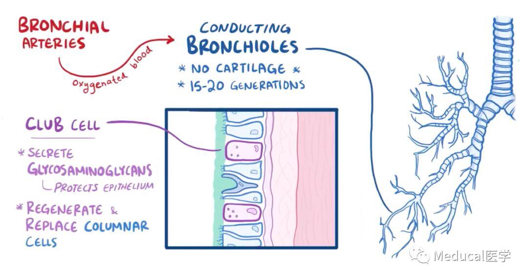
After the first three generations of bronchi, however, the airways become more narrow, called bronchioles “little bronchi”, and these can stay open without the need for cartilage. Air is conducted through smaller and smaller bronchioles for about 15-20 generations, and collectively they’re known as conducting bronchioles. Now the walls of the conducting bronchioles are similarly lined by ciliated columnar cells and mucus secreting goblet cells, as well as a new cell type called club cells because they look like tiny clubs. These club cells secrete glycosaminoglycans which is a material that protects the bronchiolar epithelium. These guys can transform into ciliated columnar cells, so they help regenerate and replace damaged ciliated columnar epithelial cells if needed. These conducting bronchioles receive oxygenated from the bronchial ateries.
肺 泡
传导性细支气管的终端是末端细支气管,之后空气通过他们进入呼吸性细支气管,这很独特,因为它们的管壁上有微小突起,称为肺泡,在肺部大约有5亿个肺泡。最终呼吸性细支气管结束于肺泡,此时气道被称为肺泡管而非呼吸细支气管。这是呼入的空气最终的目的地。
The last part of the conducting bronchioles are the terminal bronchioles, and then after that air gets to the respiratory bronchioles, which are unique because they have tiny outpouchings that bud off of their walls. These outpouchings are called alveoli, and there are about 500 million of them within the lungs. Eventually the respiratory bronchioles ends when there are no noting but alveoli, and at that point the airway is called an alveolar duct rather than a respiratory bronchiole. This is the final destination of the inhaled air.
肺泡壁与支气管壁结构完全不同:没有纤毛和平滑肌,相反管壁内有大量薄层上皮细胞,称为肺泡细胞。大部分为I型肺泡细胞,还有一些为II型肺泡细胞,可分泌一种表面活性剂,这有助于降低肺泡内的表面张力,并保持它们的开放。与棒状细胞类似,II型肺泡细胞可转化成I型肺泡细胞,替代和修复受损的细胞。
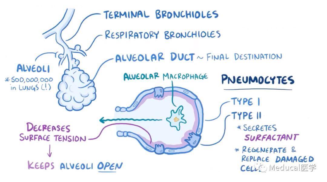
The alveolar wall has a completely different structure from the bronchioles – there are no cilia or smooth muscle, and instead the wall is lined by thin epithelial cells called pneumocytes. Most are regular pneumocytes called type I pnenumocytes, but some, called type II pneumocytes, have the ability to secrete a substance called surfactant, which helps decrease the surface tension within the alveoli and keeps them open. Like the club cells, the type II pneumocytes are capable of transforming into type I pneumocyte, so they can also help regenerate and replace damaged cells.
最后,如果极小的颗粒被吸入肺泡深处,肺泡里的巨噬细胞会吞了它,并将它移到传导性细支气管里启动粘液纤毛活动梯,送到喉部被咳出或者是被咽下去。移除异物后,被吸入到肺泡的空气周围围绕的大多是I型肺泡细胞。在肺泡细胞的另一边是内皮细胞,含有丰富的毛细血管壁——我们的血液就在这里。
Finally, if a tiny particle ever makes it deep into the lungs, there are alveolar macrophages that can gobbles it up and then physically move up to the conducting bronchioles where they can ride the mucociliary escalators all the way up to the pharynx to be either coughed up or swallowed down. Free from particles, the inhaled air is now in the alveolus surrounded by mostly type I pneumocytes. On the other side of the pneumocytes are endothelial cells that line the capillary walls – which is where that sweet sweet blood is.
然而,来自肺动脉的血液不含氧。肺泡细胞和毛细血管被一层称为基底膜的蛋白质层粘合在一起。所以,肺泡壁、基底膜和毛细血管壁将空气和血液分离,这个结构叫气血屏障。
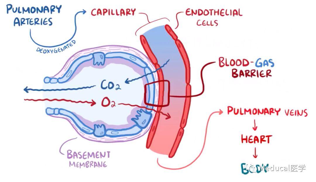
This time, though, that blood comes from the pulmonary ateries, carrying deoxygenated. The pneumocytes and capillaries are glued together with a protein layer called basement membrane. So the alveolar wall, the basement membrane, and the capillary wall is really all that separates the air from the blood, and this called the blood-gas barrier.
此刻,二氧化碳从不含氧的血液中扩散出来,进入肺泡,之后被呼出。每次呼吸,氧气都会进入肺泡,通过自由扩散进入血液。富含氧气的血液进入肺静脉、心脏最后到全身组织。
At this point, carbon dioxide diffuses out from the deoxygenated blood and into the air of the alveoli, which then gets breathed out. And with each breath in, oxygen enters the alveoli and freely diffuses into the blood. That freshly oxygenated blood then heads off to the pulmonary veins, the heart, and then to the body’s tissues.
小 结
呼吸系统主管气体交换。空气中的氧气被吸入,经过咽部、喉部、气管、上呼吸道、传导性细支气管、呼吸性细支气管、肺泡,最后是毛细血管,进入全身组织。二氧化碳会走相反的路径,最终被呼出体外。
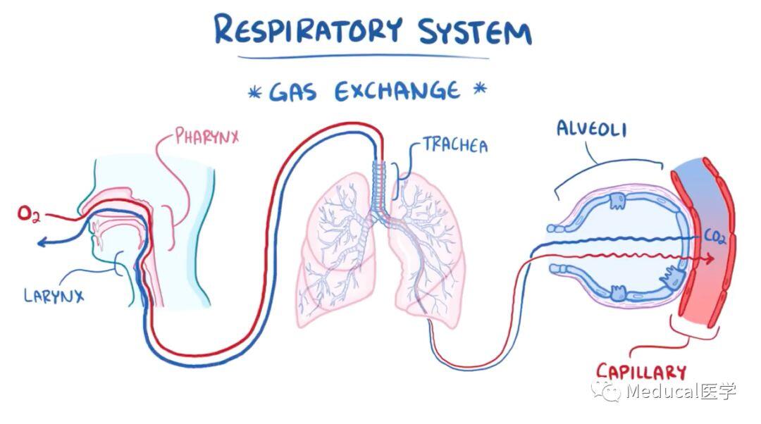
All right, as a quick recap, the respiratory system facilitates gas-exchange. Oxygen in the air is inhaled and makes it’s way through the pharynx, larynx, trachea, large upper airways, conducting bronchioles, respiratory bronchioles, the alveoli, and finally the capillary to be sent to the body’s tissue. The carbon dioxide makes the reverse journey to eventually be exhaled into the world.
Osmosis玩呀
https://www.osmosis.org/invite/gL9HB3x
